
Gigas Larvae Day 3
Counting larvae takes a while
Before I get into that, a quick rundown of other things I did today.
- Recorded probe numbers in each larval bucket
- 1: SN6 Amb pH A
- 2: SN6 Low pH B
- 3: SN6 Amb B
- 5: NF6 Amb B
- 6: NF10 Low pH A (Found out later in the day that this probe stopped working in May)
- 9: SN10 Low pH A
- 10: SN10 Amb A
- 11: SN6 Low pH A
- 12: NF6 Amb A
- 12: NF10 Amb B
- 15: NF6 Low pH B
- 17: NF10 Low pH B
- 19: SN10 Low B
- 21: NF6 Low pH A
- 23: NF10 Amb A
- Recorded which bucket was in which tote

Figure 1. Tote layout. Totes go from 1 to 9, left to right.
- Did not record morning temperatures
- HOBOs already in tanks, no need
- Measured morning food presence
- Very dense! I think this is because I fed in the afternoon on Tuesday, so the larvae didn’t have a full 24 hours to really feed down

Figure 2. Color of larval tanks in the morning, before daily feeding.
- Steven rigged up the AVTECH in the SeaLab, so I now have 5 probes to work with
- He also started messing around with the OA system set-up
- Moved immersion heater probes to inside larval bucket, instead of in tote
- Counted heaters
- 8 new from Roberts Lab, 1 not being used right now
- 2 old heaters (one I ordered, one I took from the lab with the Farenheight controller)
- Gray heater borrowed from rick
- Fed larvae
Now to the main event: imaging and counting! I counted all of the larvae in Plate 1 collected yesterday. It took me much longer than I expected, mainly because I was trying to figure out how to work the dissecting scope while also taking pictures for future use. My count data can be found here.
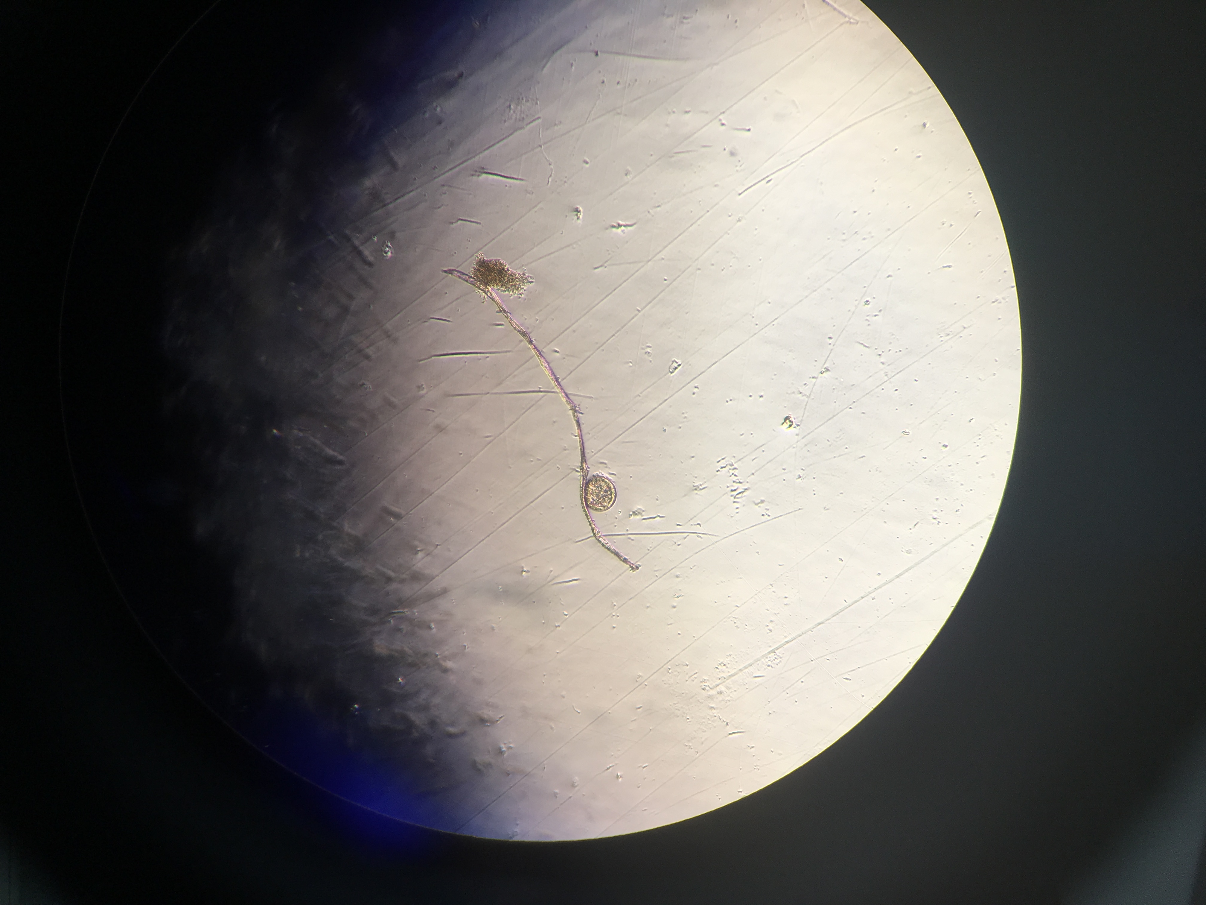
There were lots of larvae that exhibited a normal D-hinge shape, but there were also some that looked like peanuts! I recorded how many of these deformed ones I saw as well.
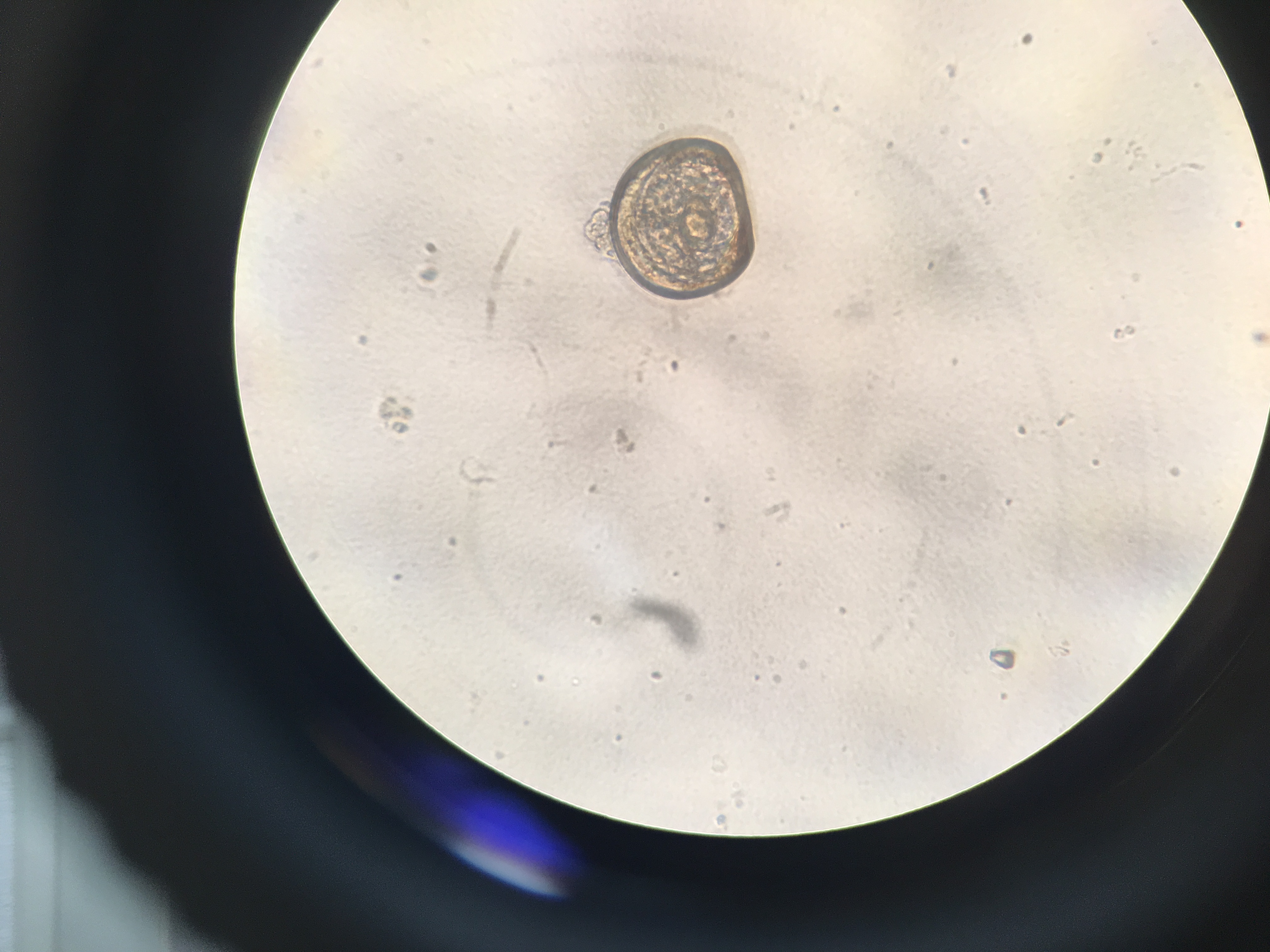
Figures 3-4. Normal D-hinge larvae shape at 40x (top) and 20x (bottom) magnification.

Figure 5. Weird larvae shape.
I’m storing all of my larvae photos and videos from today here. I’m cataloging each day in a different folder, and labelling which bucket the larvae are from in the image name.
For tomorrow:
- Screen with 60 and 48 micron screens
- Sample and count
- Save samples in -80ºC freezer
- Finish imaging Plate 2 and 3
Since you made it through this lab post, here are some easy links to larvae videos. Both are from bucket 8: